Argon »
PDB 1c66-7q38 »
6q8q »
Argon in PDB 6q8q: Bovine Insulin Under 2 Kbar of Argon
Protein crystallography data
The structure of Bovine Insulin Under 2 Kbar of Argon, PDB code: 6q8q
was solved by
T.Prange,
P.Carpentier,
with X-Ray Crystallography technique. A brief refinement statistics is given in the table below:
| Resolution Low / High (Å) | 38.81 / 2.00 |
| Space group | I 21 3 |
| Cell size a, b, c (Å), α, β, γ (°) | 77.559, 77.559, 77.559, 90.00, 90.00, 90.00 |
| R / Rfree (%) | 18.2 / 19.9 |
Argon Binding Sites:
The binding sites of Argon atom in the Bovine Insulin Under 2 Kbar of Argon
(pdb code 6q8q). This binding sites where shown within
5.0 Angstroms radius around Argon atom.
In total only one binding site of Argon was determined in the Bovine Insulin Under 2 Kbar of Argon, PDB code: 6q8q:
In total only one binding site of Argon was determined in the Bovine Insulin Under 2 Kbar of Argon, PDB code: 6q8q:
Argon binding site 1 out of 1 in 6q8q
Go back to
Argon binding site 1 out
of 1 in the Bovine Insulin Under 2 Kbar of Argon
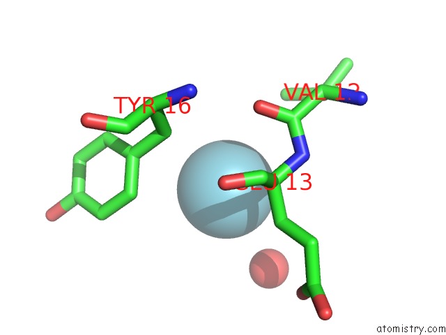
Mono view
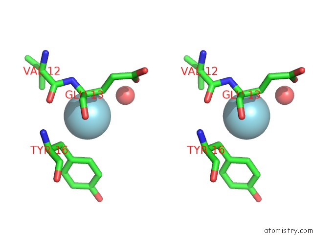
Stereo pair view
Mono view
Stereo pair view
A full contact list of Argon with other atoms in the Ar binding
site number 1 of Bovine Insulin Under 2 Kbar of Argon within 5.0Å range:
|
Reference:
T.Prange,
P.Carpentier.
Argon-Labelling Protein Crystals By High Pressure Cooling at 2 Kbar To Be Published.
Page generated: Sun Jul 6 22:43:53 2025
Last articles
Cl in 8D6BCl in 8D69
Cl in 8D62
Cl in 8D60
Cl in 8D61
Cl in 8D5Z
Cl in 8D5X
Cl in 8D5U
Cl in 8D5W
Cl in 8D5T