Argon »
PDB 1c66-7q38 »
7q38 »
Argon in PDB 7q38: Crystal Structure of the Mutant Bacteriorhodopsin Pressurized with Argon
Protein crystallography data
The structure of Crystal Structure of the Mutant Bacteriorhodopsin Pressurized with Argon, PDB code: 7q38
was solved by
I.Melnikov,
M.Rulev,
R.Astashkin,
K.Kovalev,
P.Carpentier,
V.Gordeliy,
A.Popov,
with X-Ray Crystallography technique. A brief refinement statistics is given in the table below:
| Resolution Low / High (Å) | 83.02 / 1.65 |
| Space group | C 2 2 2 |
| Cell size a, b, c (Å), α, β, γ (°) | 115.657, 119.245, 36.393, 90, 90, 90 |
| R / Rfree (%) | 16.2 / 20.5 |
Argon Binding Sites:
Pages:
>>> Page 1 <<< Page 2, Binding sites: 11 - 20; Page 3, Binding sites: 21 - 30; Page 4, Binding sites: 31 - 40; Page 5, Binding sites: 41 - 47;Binding sites:
The binding sites of Argon atom in the Crystal Structure of the Mutant Bacteriorhodopsin Pressurized with Argon (pdb code 7q38). This binding sites where shown within 5.0 Angstroms radius around Argon atom.In total 47 binding sites of Argon where determined in the Crystal Structure of the Mutant Bacteriorhodopsin Pressurized with Argon, PDB code: 7q38:
Jump to Argon binding site number: 1; 2; 3; 4; 5; 6; 7; 8; 9; 10;
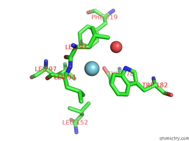
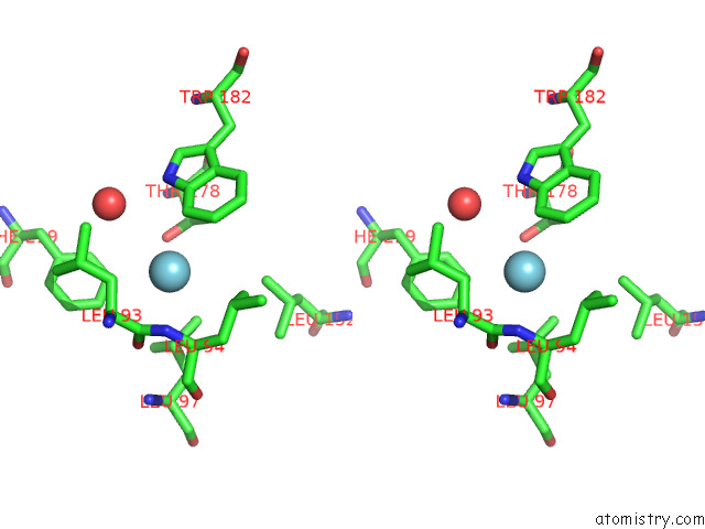
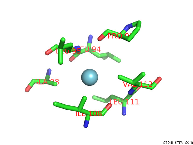
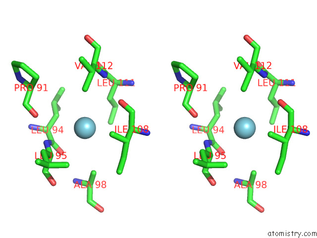
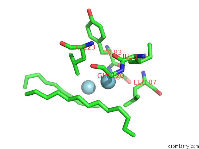
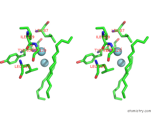
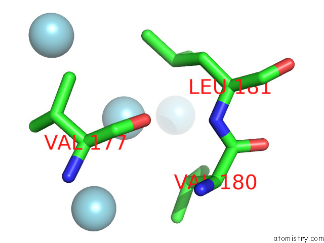
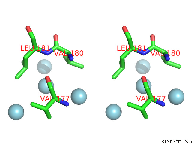
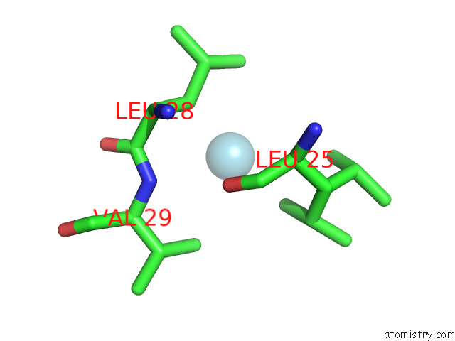
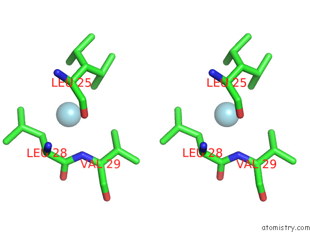
Argon binding site 1 out of 47 in 7q38
Go back to
Argon binding site 1 out
of 47 in the Crystal Structure of the Mutant Bacteriorhodopsin Pressurized with Argon
Mono view
Stereo pair view
Mono view
Stereo pair view
A full contact list of Argon with other atoms in the Ar binding
site number 1 of Crystal Structure of the Mutant Bacteriorhodopsin Pressurized with Argon within 5.0Å range:
|
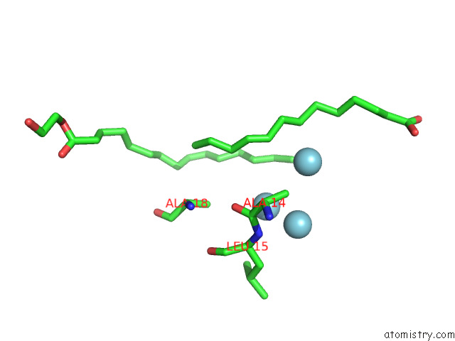
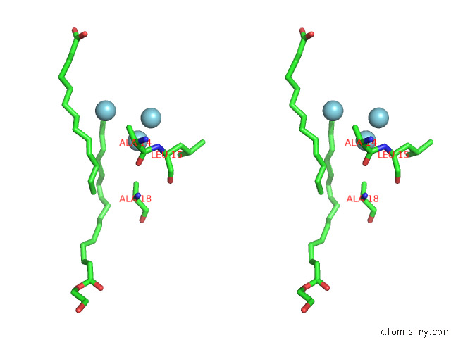
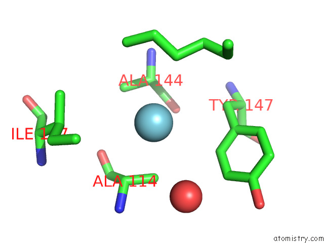
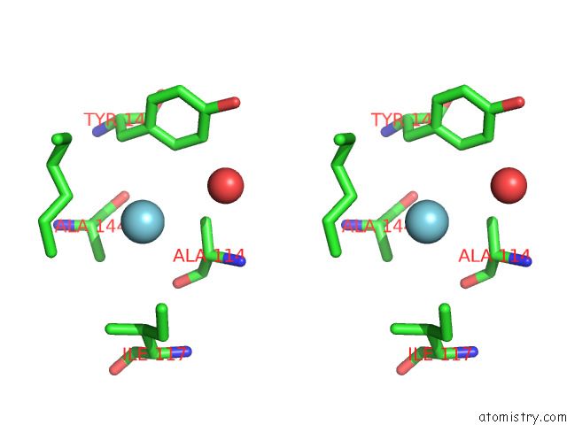
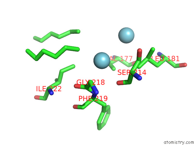
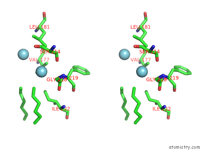
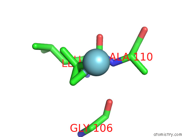
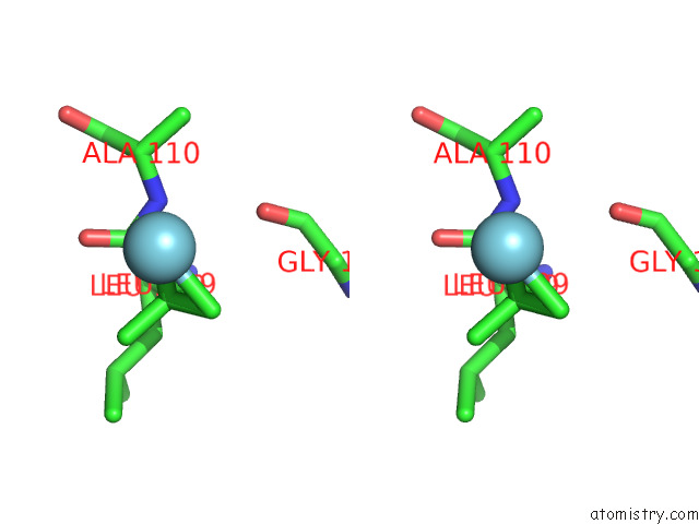
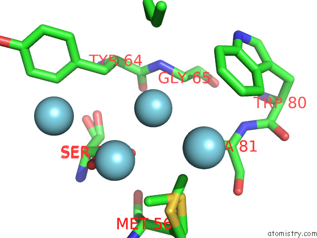
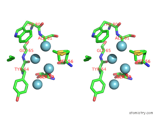
Argon binding site 2 out of 47 in 7q38
Go back to
Argon binding site 2 out
of 47 in the Crystal Structure of the Mutant Bacteriorhodopsin Pressurized with Argon
Mono view
Stereo pair view
Mono view
Stereo pair view
A full contact list of Argon with other atoms in the Ar binding
site number 2 of Crystal Structure of the Mutant Bacteriorhodopsin Pressurized with Argon within 5.0Å range:
|
Argon binding site 3 out of 47 in 7q38
Go back to
Argon binding site 3 out
of 47 in the Crystal Structure of the Mutant Bacteriorhodopsin Pressurized with Argon
Mono view
Stereo pair view
Mono view
Stereo pair view
A full contact list of Argon with other atoms in the Ar binding
site number 3 of Crystal Structure of the Mutant Bacteriorhodopsin Pressurized with Argon within 5.0Å range:
|
Argon binding site 4 out of 47 in 7q38
Go back to
Argon binding site 4 out
of 47 in the Crystal Structure of the Mutant Bacteriorhodopsin Pressurized with Argon
Mono view
Stereo pair view
Mono view
Stereo pair view
A full contact list of Argon with other atoms in the Ar binding
site number 4 of Crystal Structure of the Mutant Bacteriorhodopsin Pressurized with Argon within 5.0Å range:
|
Argon binding site 5 out of 47 in 7q38
Go back to
Argon binding site 5 out
of 47 in the Crystal Structure of the Mutant Bacteriorhodopsin Pressurized with Argon
Mono view
Stereo pair view
Mono view
Stereo pair view
A full contact list of Argon with other atoms in the Ar binding
site number 5 of Crystal Structure of the Mutant Bacteriorhodopsin Pressurized with Argon within 5.0Å range:
|
Argon binding site 6 out of 47 in 7q38
Go back to
Argon binding site 6 out
of 47 in the Crystal Structure of the Mutant Bacteriorhodopsin Pressurized with Argon
Mono view
Stereo pair view
Mono view
Stereo pair view
A full contact list of Argon with other atoms in the Ar binding
site number 6 of Crystal Structure of the Mutant Bacteriorhodopsin Pressurized with Argon within 5.0Å range:
|
Argon binding site 7 out of 47 in 7q38
Go back to
Argon binding site 7 out
of 47 in the Crystal Structure of the Mutant Bacteriorhodopsin Pressurized with Argon
Mono view
Stereo pair view
Mono view
Stereo pair view
A full contact list of Argon with other atoms in the Ar binding
site number 7 of Crystal Structure of the Mutant Bacteriorhodopsin Pressurized with Argon within 5.0Å range:
|
Argon binding site 8 out of 47 in 7q38
Go back to
Argon binding site 8 out
of 47 in the Crystal Structure of the Mutant Bacteriorhodopsin Pressurized with Argon
Mono view
Stereo pair view
Mono view
Stereo pair view
A full contact list of Argon with other atoms in the Ar binding
site number 8 of Crystal Structure of the Mutant Bacteriorhodopsin Pressurized with Argon within 5.0Å range:
|
Argon binding site 9 out of 47 in 7q38
Go back to
Argon binding site 9 out
of 47 in the Crystal Structure of the Mutant Bacteriorhodopsin Pressurized with Argon
Mono view
Stereo pair view
Mono view
Stereo pair view
A full contact list of Argon with other atoms in the Ar binding
site number 9 of Crystal Structure of the Mutant Bacteriorhodopsin Pressurized with Argon within 5.0Å range:
|
Argon binding site 10 out of 47 in 7q38
Go back to
Argon binding site 10 out
of 47 in the Crystal Structure of the Mutant Bacteriorhodopsin Pressurized with Argon
Mono view
Stereo pair view
Mono view
Stereo pair view
A full contact list of Argon with other atoms in the Ar binding
site number 10 of Crystal Structure of the Mutant Bacteriorhodopsin Pressurized with Argon within 5.0Å range:
|
Reference:
I.Melnikov,
P.Orekhov,
M.Rulev,
K.Kovalev,
R.Astashkin,
D.Bratanov,
Y.Ryzhykau,
T.Balandin,
S.Bukhdruker,
I.Okhrimenko,
V.Borshchevskiy,
G.Bourenkov,
C.Mueller-Dieckmann,
P.Van Der Linden,
P.Carpentier,
G.Leonard,
V.Gordeliy,
A.Popov.
High-Pressure Crystallography Shows Noble Gas Intervention Into Protein-Lipid Interaction and Suggests A Model For Anaesthetic Action. Commun Biol V. 5 360 2022.
ISSN: ESSN 2399-3642
PubMed: 35422073
DOI: 10.1038/S42003-022-03233-Y
Page generated: Wed Jul 10 10:50:51 2024
ISSN: ESSN 2399-3642
PubMed: 35422073
DOI: 10.1038/S42003-022-03233-Y
Last articles
Zn in 9MJ5Zn in 9HNW
Zn in 9G0L
Zn in 9FNE
Zn in 9DZN
Zn in 9E0I
Zn in 9D32
Zn in 9DAK
Zn in 8ZXC
Zn in 8ZUF